Lab Publications and Conferences
Peer Reviewed Publications
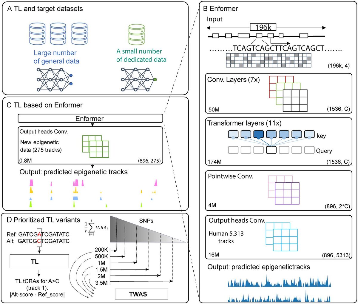
38. Tissue-specific Transfer Learning Improves Functional Variant and Therapeutic Target Discoveries in Breast and Prostate Cancer
DNA foundation models trained on large-scale genomic and epigenetic datasets have shown promise for regulatory variant interpretation, yet their application to tissue-specific contexts remain limited. Here, we present a transfer learning (TL) framework to adapt Enformer, a deep neural network trained on 5,313 multi-omics tracks, to breast and prostate cancer using 275 and 357 tissue-specific transcription factor (TF) ChIP–seq tracks, respectively. We computed tissue-specific cis-regulatory activity (tCRA) scores for millions of single-nucleotide variants (SNVs) in genome-wide association study (GWAS) datasets and prioritized high-impact SNV subsets (1M, 1.5M, and 2M). These TL-prioritized variants demonstrated consistently greater enrichment in tissue-specific enhancers, cancer GWAS risk variants, and ClinVar pathogenic variants compared to the original Enformer model. Transcriptome-wide association studies (TWAS) using TL-based SNVs identified more cancer-relevant genes, many of which exhibited functional essentiality (DepMap), therapeutic tractability (drug databases), and disease relevance (DisGeNET). Notably, TL models outperformed the base model in identifying genes enriched for drug targets and clinically relevant disease associations. Our results show that TL-derived tCRA scores enhance regulatory variant prioritization and improve susceptibility gene discovery in a tissue-specific manner. Our study provides a generalizable framework for tailoring foundation models to disease-relevant contexts, with implications for variant interpretation, therapeutic target discovery, and precision medicine.
37. A Multi-Site Harmonization of Computed Tomography Perfusion Maps using Scaling Factors and ComBat
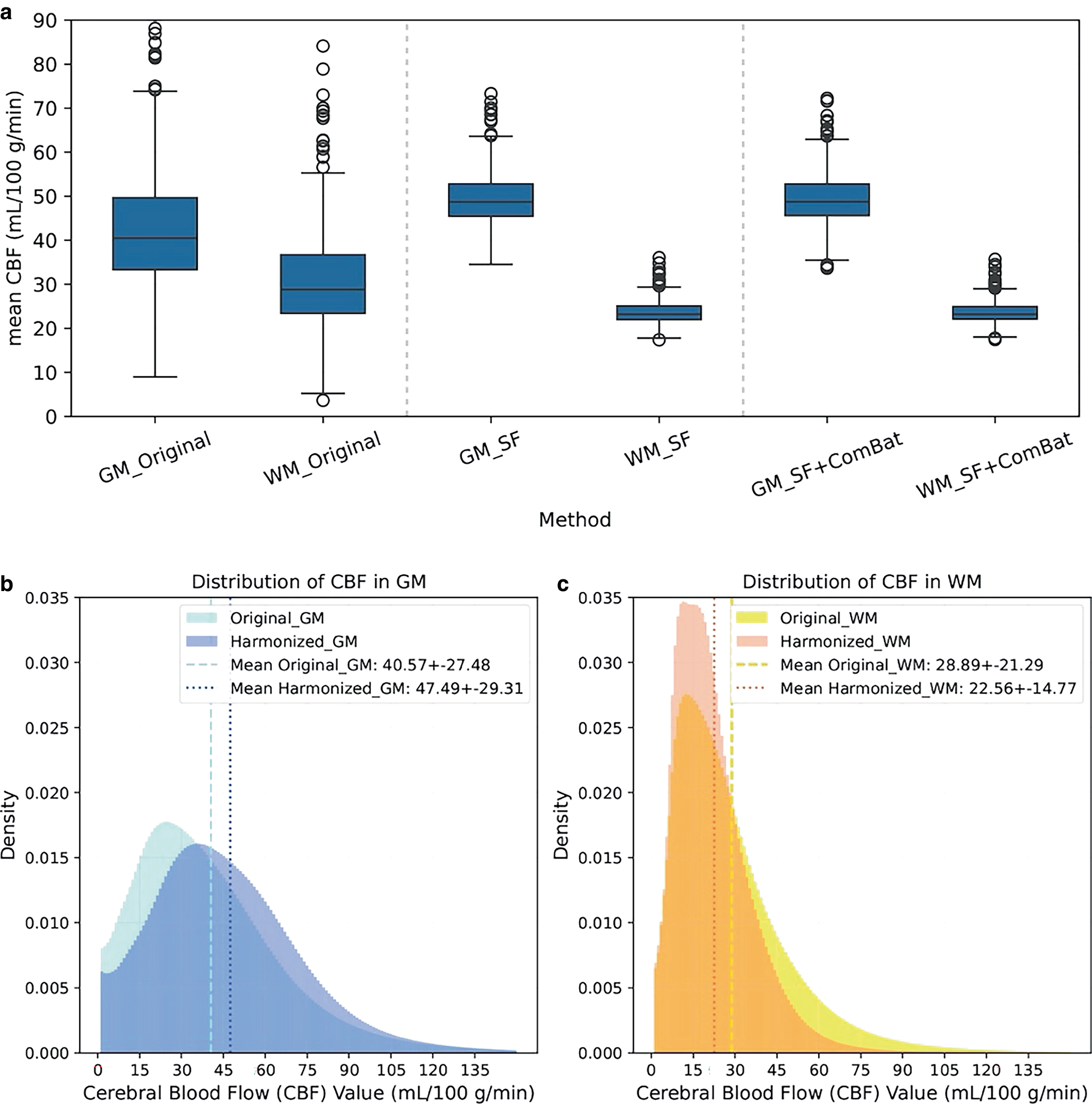
Computed tomography perfusion (CTP)-derived cerebral blood flow (CBF) maps are widely used to study infarct volume and growth. Multi-site CTPs often have site biases that can alter thresholds and increase errors. However, the clinical impact of inter-site variability on infarct quantification is unclear. We examined this variability and developed an automated harmonization pipeline for multi-site CTP datasets to mitigate infarct assessment errors. We analyzed 741 CTP cases from the Alteplase compared to Tenecteplase trial across eight sites. CBFs were registered and segmented into grey-matter/white-matter (GM/WM). We scaled mean GM/WM CBFs into physiological ranges and applied ComBat, an empirical Bayesian method, to reduce inter-site bias. We assessed clinical utility by comparing infarct-core volumes from original versus harmonized maps with a relative-CBF-based reference, and comparing large-core infarct identification using CBF-based volume thresholds. The original mean CBF deviated from physiological range with high variance and significant inter-site differences. After applying the scaling factor, the mean CBF (GM/WM) shifted to physiological ranges (49.3 ± 6.0/23.5 ± 2.5 ml/100g/min), and ComBat effectively eliminated inter-site differences. In 199 cases with complete reference data for clinical evaluation, harmonization decreased mean absolute error for infarct volume by 39% (12.7 to 7.7 ml), improved agreement (bias −7.9 to −5.8 ml; narrower 95%CI), and increased accuracy of large-core classification at a 60 ml-threshold by 5.0% (net reclassification improvement:0.310, 95%CI:0.036–0.603). Our harmonization pipeline rescaled multi-site CBF maps to physiological range, reducing inter-site variability and errors in infarct core estimation and large-core classification. Multi-site harmonization should become standard in CTP datasets to avoid erroneous conclusions about infarct size and growth.
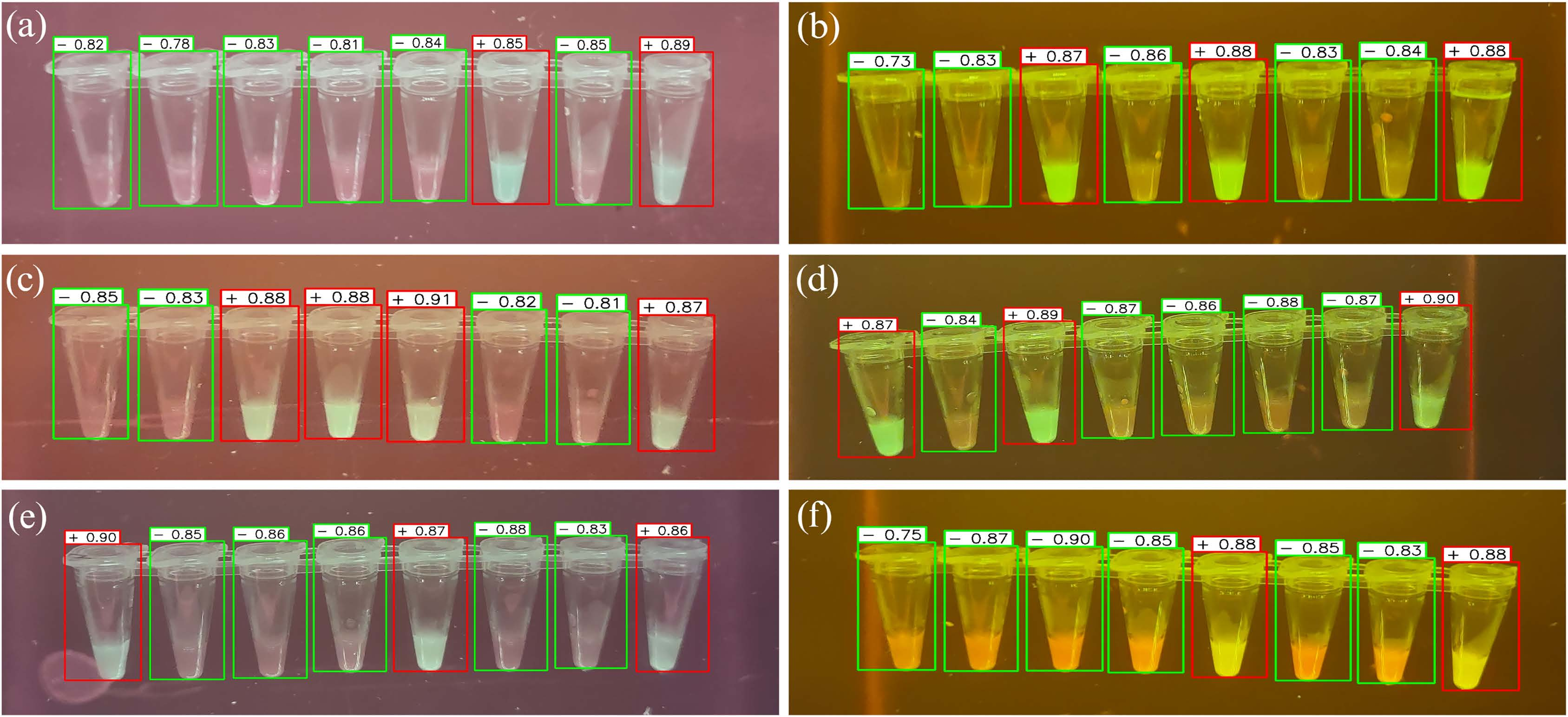
36. You Only Look Once (YOLO) Based Machine Learning Algorithm for Real-Time Detection of Loop-Mediated Isothermal Amplification (LAMP) Diagnostics
Loop-mediated isothermal amplification (LAMP) is a widely used rapid and affordable molecular DNA amplification method with minimal resource requirements. However, visual interpretation of results is subjective and prone to errors, leading to potential false-positive and negative results. To address this limitation, a machine-learning approach is proposed for automated LAMP classification based on digital images. The approach utilizes You Only Look Once (YOLOv8), a fast and robust object detection algorithm to locate and classify tubes within LAMP images, enabling automated categorization as positive or negative. The trained model achieved a high overall accuracy of 97.4% in classifying LAMP images into positive or negative on the test set. Additionally, the approach had a 95.3% precision and 96.8% recall for positive cases and 93.3% precision and 95.8% recall for negative cases, demonstrating its potential for real-time LAMP diagnosis and enhanced assay performance. This project demonstrated platform suitability for real-time testing, offering an easy operation and rapid results.
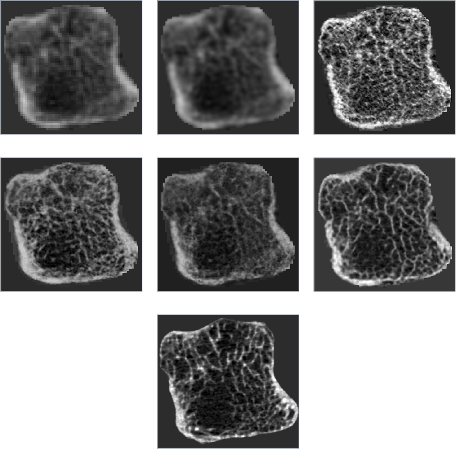
35. CL-GAN: A Progressive Curriculum Learning Approach for Bone CT Super-Resolution
Cone-beam computed tomography (CBCT) provides rapid, low-dose imaging with large anatomical coverage but lacks the resolution required for detailed bone assessment in rheumatoid arthritis (RA). Enhancing CBCT to match high-resolution peripheral quantitative CT (HR-pQCT) is challenging due to differences in resolution, noise, and artifact profiles, causing standard super-resolution approaches to struggle in training with instability. We propose CL-GAN (curriculum learning generative adversarial network) to train a Cycle-Consistent GAN for CBCT super-resolution. Training is structured into four stages, beginning with paired synthetic mappings and gradually introducing unpaired real CBCT and HR-pQCT images from healthy and RA-affected joints. Each stage progressively increases task difficulty to bridge domain gaps stably. Performance was evaluated using image quality metrics, trabecular bone morphometry, and blinded review by an experienced reviewer. Repeated measures ANOVA and post-hoc paired t-tests assessed significance across stages. Progressive training led to consistent improvements in image quality and trabecular bone metric accuracy, with all gains statistically significant (p<0.001). In blinded review, two reviewers detected all RA erosions while one missed one erosion, and all reviewers reported difficulty distinguishing enhanced CBCT from HR-pQCT. An ablation study showed that skipping curriculum stages impaired image quality, highlighting the importance of gradual domain adaptation. This study demonstrates that curriculum learning enables stable and effective training of a super-resolution algorithm for CBCT images in the context of RA. By enhancing image quality and structural interpretability, the proposed framework increases the amount of relevant information that can be extracted from CBCT, supporting its utility in RA analysis and research.
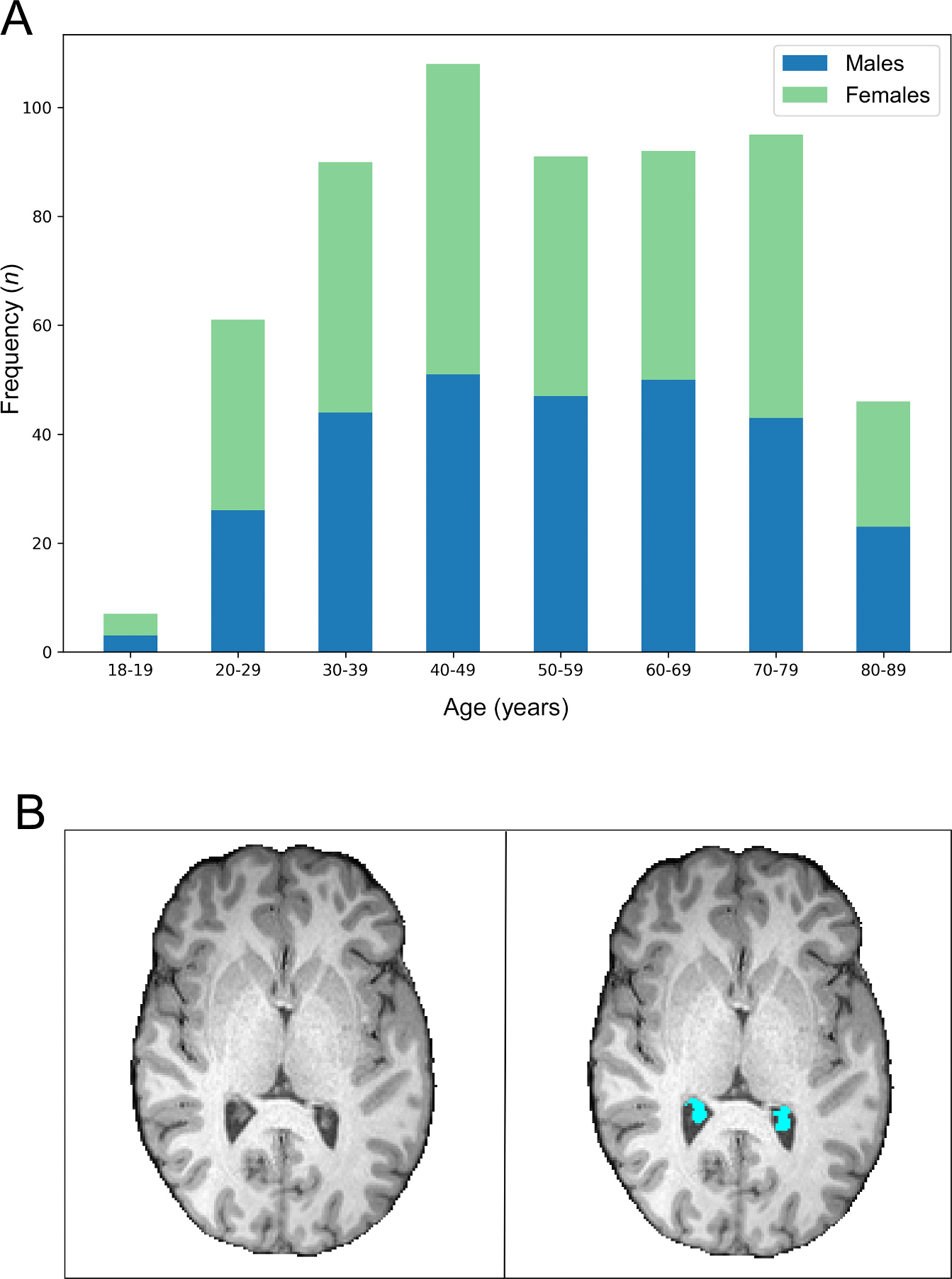
34. Choroid Plexus Volume and its Association with Cognitive Performance Across the Lifespan: Links to Sleep Quality and Healthy Brain Aging
The choroid plexus (ChP) is implicated in inflammation and supports the clearance of waste byproducts, particularly those related to the pathogenesis of Alzheimer’s disease. Increases in ChP volume have been associated with older age and cognitive decline in both clinical and healthy cohorts. However, the clearance of waste products in the brain is also related to sleep, and sleep quality may contribute to ChP dysfunction and cognitive decline. In the present work, it was therefore hypothesized that the association between age and cognitive performance is mediated by ChP volume, however this is conditional on sleep quality. A moderated-mediation model was tested on a sample ( N = 590) of healthy adults aged 18–87 years from the Cambridge Centre for Ageing and Neuroscience (Cam-CAN). Results showed that the relationship between increasing age and decreased cognitive performance was partially mediated by ChP volume, however, this was not conditional on sleep quality. A moderation analysis indicated that the relationship between ChP volume and cognitive performance was moderated by age, with ChP enlargement associated with worse cognitive performance in participants older than 62 years. In participants younger than 62 years, sleep duration was associated with cognitive performance, but ChP volume was not. These findings provide support for the sensitivity of ChP volume to cognitive performance in older adults. • In healthy adults, increasing age was associated with decreasing cognitive performance. • ChP volume partially mediated this relationship between age and cognitive performance. • Sleep quality predicted cognitive performance in younger but not older adults. • ChP volume predicted cognitive performance in older but not younger adults.
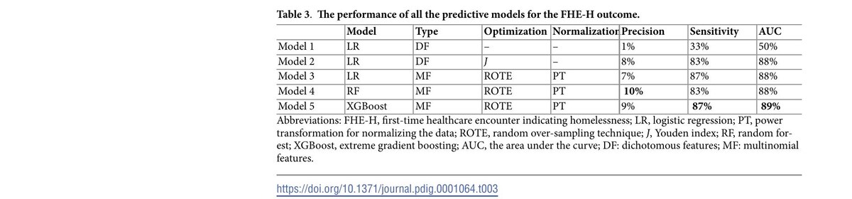
33. Risk factors and predictive performance for first healthcare encounters indicating homelessness and police interactions using administrative data among residents of Calgary, Alberta, Canada who are diagnosed with addiction or mental health conditions
Individuals diagnosed with addiction or mental health (AMH) conditions are more likely to experience potentially adverse outcomes of homelessness. Despite their link to later outcomes, research on initial episodes of AMH outcomes is limited. This study aims to use administrative data to identify the factors associated with the first healthcare encounters with indicators of homelessness (FHE-H) for individuals diagnosed with AMH. We assessed logistic regression and compared its performance with machine learning models, including random forests and extreme gradient boosting (XGBoost). We conducted a retrospective cohort study linking several administrative datasets for 232,253 individuals with Alberta health insurance in Calgary, Canada, who were aged between 18 and 65 and diagnosed with AMH between April 1, 2013, and March 31, 2018. We assessed outcomes in two years following cohort entry. Individuals with episodes of FHE-H (2,606 individuals) before the index date were excluded. Multivariable logistic regression models were used to identify factors associated with outcomes by estimating adjusted odds ratios (AORs) with 95% confidence intervals. Among 229,647 individuals diagnosed with AMH, 1,886 (0.82%) experienced FHE-H during the follow-up period. Mental health emergency visits (AORs=5.28 [95% CI: 4.41, 6.33]), substance misuse (AORs=3.87 [95% CI: 3.28, 4.56], substance use disorders (AORs=2.03 [95% CI: 1.64, 2.50]), and male sex (AORs=1.28 [95% CI: 1.14, 1.44]) were associated with FHE-H. XGBoost performance improved over logistic regression, with increases in area under the curve (AUC) by 1% and precision by 2%. Overall, several AMH features were associated with FHE-H, and machine learning models outperformed logistic regression, although to a small degree.
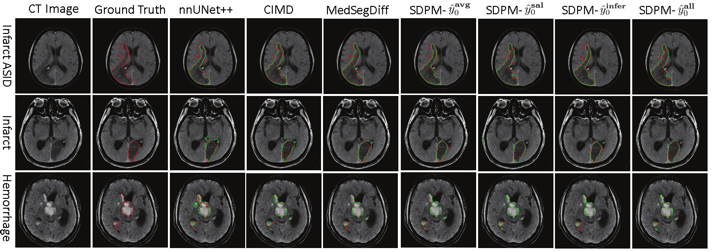
32. Synchronous Image-Label Diffusion Probability Model with Application to Stroke Lesion Segmentation on Non-contrast CT
The stroke lesion volume is a key radiologic measurement for assessing the prognosis of acute ischemic stroke (AIS) patients, which is challenging to be automatically measured on noncontrast CT (NCCT) scans. Recent diffusion probabilistic models (DPMs) in the domain of image generation have shown potentials of being used for lesion volume segmentation on medical images. In this article, a novel synchronous image-label diffusion probability model (SDPM) is proposed for stroke lesion segmentation on NCCT using a dual-Markov diffusion process with shared noise. The proposed SDPM is fully based on a generative latent variable model (LVM), offering a probabilistic elaboration from stem to stem. To fit into our segmentation tasks using the strength from generation models, we develop the architecture of the network where an additional net-stream, parallel with a noise prediction stream, is introduced to obtain the initial label estimates with noise for efficiently inferring the final labels. By optimizing the specified variational boundaries, the trained model can infer the final label estimates given the input images at any scale of time in four different label-inference methods, which gives more flexibility to the proposed SDPM. The proposed model was assessed on three stroke lesion datasets including one public and two private datasets. Compared with several U-Net, transformer, and DPM-based segmentation methods, our proposed SDPM model is able to achieve the state-of-the-art accuracy.
31. Physiological Confounds in BOLD-fMRI and their Correction
Functional magnetic resonance imaging (fMRI) has opened new frontiers in neuroscience by instrumentally driving our understanding of brain function and development. Despite its substantial successes, fMRI studies persistently encounter obstacles stemming from inherent, unavoidable physiological confounds. The adverse effects of these confounds are especially noticeable with higher magnetic fields, which have been gaining momentum in fMRI experiments. This review focuses on the four major physiological confounds impacting fMRI studies: low‐frequency fluctuations in both breathing depth and rate, low‐frequency fluctuations in the heart rate, thoracic movements, and cardiac pulsatility. Over the past three decades, numerous correction techniques have emerged to address these challenges. Correction methods have effectively enhanced the detection of task‐activated voxels and minimized the occurrence of false positives and false negatives in functional connectivity studies. While confound correction methods have merit, they also have certain limitations. For instance, model‐based approaches require externally recorded physiological data that is often unavailable in fMRI studies. Methods reliant on independent component analysis, on the other hand, need prior knowledge about the number of components. Machine learning techniques, although showing potential, are still in the early stages of development and require additional validation. This article reviews the mechanics of physiological confound correction methods, scrutinizes their performance and limitations, and discusses their impact on fMRI studies.

30. Automatic Matching Algorithms to Increase Enrolment in Stroke Clinical Trials: A Proof-of-Concept Study
Background: Clinical trials often struggle to recruit enough participants, with only 10% of eligible patients enrolling. This is concerning for conditions like stroke, where timely decision-making is crucial. Frontline clinicians typically screen patients manually, but this approach can be overwhelming and lead to many eligible patients being overlooked. Methods: To address the problem of efficient and inclusive screening for trials, we developed a matching algorithm using imaging and clinical variables gathered as part of the AcT trial (NCT03889249) to automatically screen patients by matching these variables with the trials’ inclusion and exclusion criteria using rule-based logic. We then used the algorithm to identify patients who could have been enrolled in six trials: EASI-TOC (NCT04261478), CATIS-ICAD (NCT04142125), CONVINCE (NCT02898610), TEMPO-2 (NCT02398656), ESCAPE-MEVO (NCT05151172), and ENDOLOW (NCT04167527). To evaluate our algorithm, we compared our findings to the number of enrollments achieved without using a matching algorithm. The algorithm’s performance was validated by comparing results with ground truth from a manual review of two clinicians. The algorithm’s ability to reduce screening time was assessed by comparing it with the average time used by study clinicians. Results: The algorithm identified more potentially eligible study candidates than the number of participants enrolled. It also showed over 90% sensitivity and specificity for all trials, and reducing screening time by over 100-fold. Conclusions: Automated matching algorithms can help clinicians quickly identify eligible patients and reduce resources needed for enrolment. Additionally, the algorithm can be modified for use in other trials and diseases.

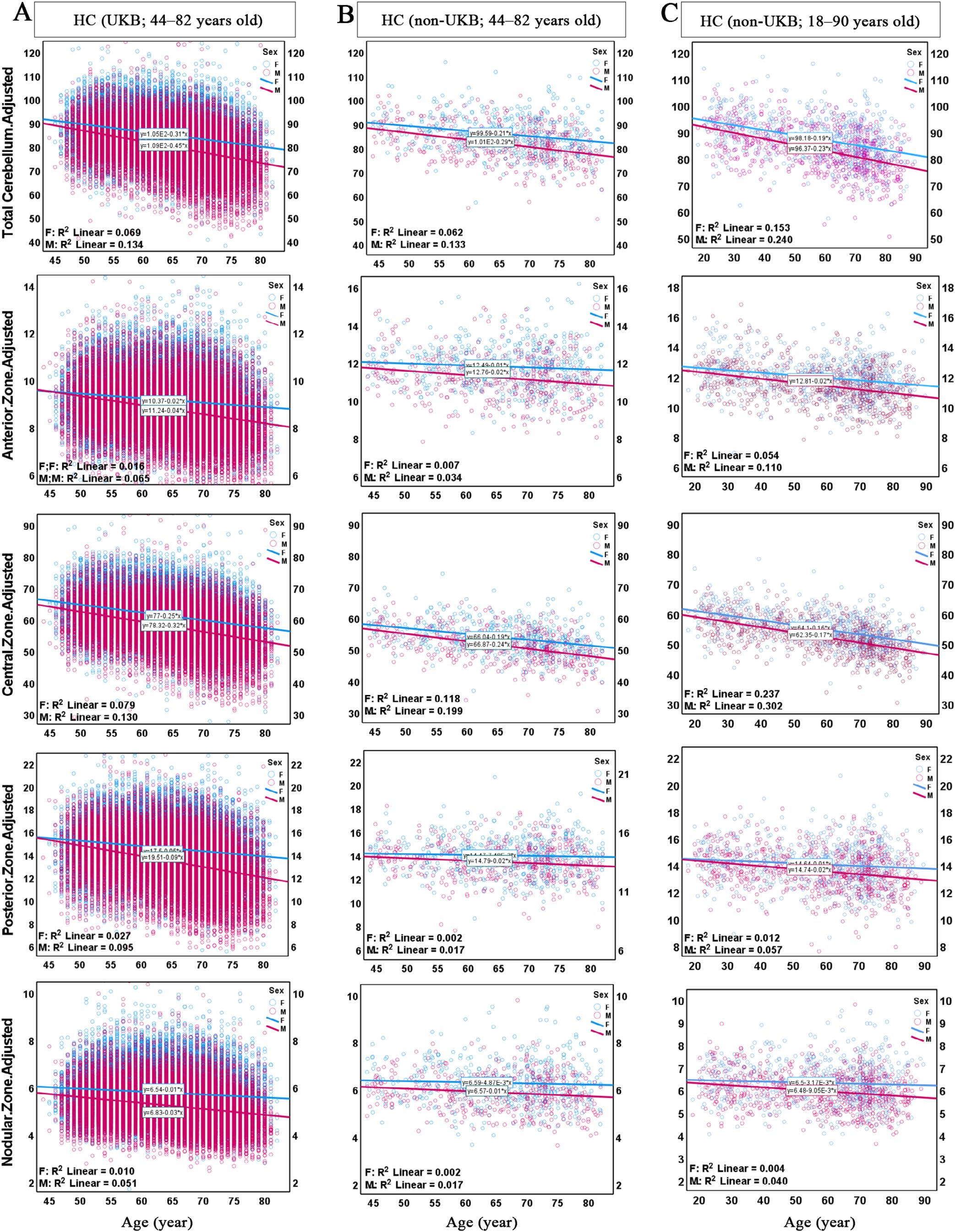
29. Volume Changes in Cerebellar Transverse Zones and Lobules: Insights from Healthy Individuals and Patients with Neurological Disorders
Cerebellar volumetric changes are intricately linked to aging, with distinct patterns across its transverse zones , the functional subdivisions characterized by unique cytoarchitectural and connectivity profiles. Despite research efforts, the cerebellar aging process in health and neurological disorders remains poorly understood. In this study, we investigated the effects of age and sex on total cerebellum, transverse zone , and lobule volumes using MRI data from over 45,000 participants compiled from six neuroimaging datasets. We also propose a framework for estimating cerebellum age as an indicator of cerebellar health. Significant age‐dependent volume reductions were observed across transverse zones , with the central zone ( CZ ; lobules VI and VII) exhibiting the steepest decline in both health and neurological disorders. This finding highlights the CZ's vulnerability to aging and its critical role in cognitive and emotional processing. We also found prominent sex differences in age‐dependent volumetric changes. Males exhibited smaller total intracranial volume (TIV)‐adjusted cerebellum volume and faster age‐dependent volume reduction than females in both health and mild cognitive impairment (MCI), Alzheimer disease (AD), and Parkinson disease (PD). In contrast, females with schizophrenia (SZ) and cocaine use disorder (CUD) revealed faster age‐dependent cerebellar volume reduction than males. Patients with MCI, AD, and PD experienced more pronounced atrophy in the posterior ( PZ ) and nodular ( NZ ) zones compared to age‐matched healthy controls, while SZ patients were characterized by a more prominent reduction in CZ . In CUD, a non‐significant volume decline was observed in all zones compared to the controls. Moreover, our framework for estimating cerebellum age revealed a notable difference in cerebellar aging between healthy individuals and neurological patients. Finally, we charted age‐dependent changes in cerebellar volume in healthy individuals, focusing on transverse zones capturing the functional subdivisions. These findings underscore the potential of cerebellar volumetric analysis as a biomarker for early detection and monitoring of neurodegenerative and neuropsychiatric disorders. Our novel approach complements and enhances MRI‐based analyses, providing essential insights into the pathogenesis of aging, neurodegeneration, and chronic neuropsychiatric conditions.
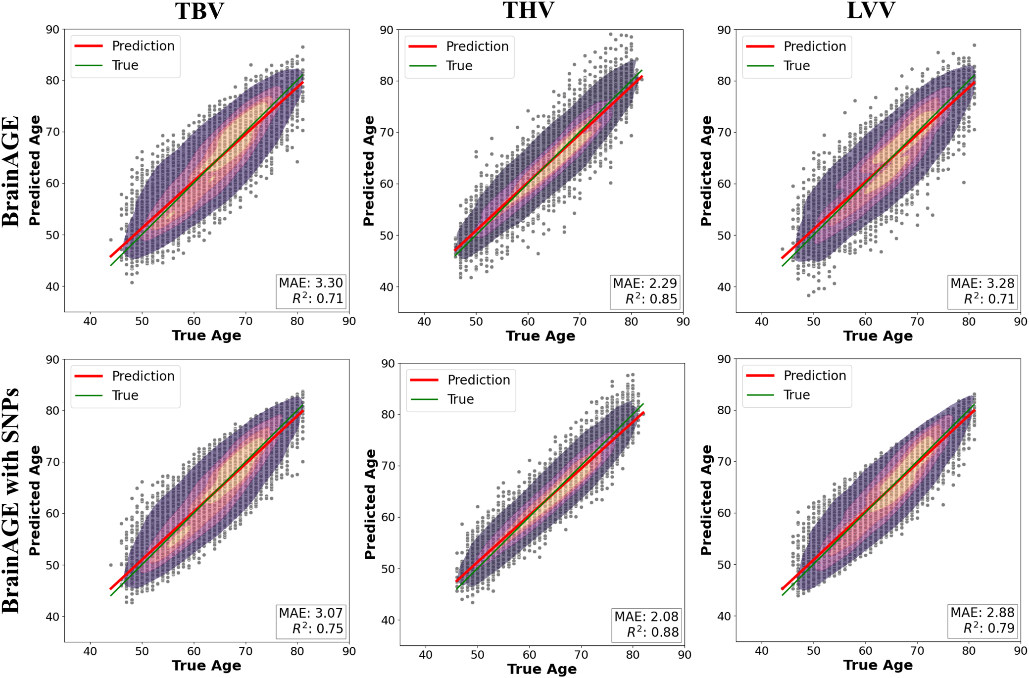
28. Integration of Genetic Information to Improve Brain Age Gap Estimation Models in the UK Biobank
Neurodegeneration occurs when the body's central nervous system becomes impaired as a person ages, which can happen at an accelerated pace. Neurodegeneration impairs quality of life, affecting essential functions, including memory and the ability to self-care. Genetics play an important role in neurodegeneration and longevity. Brain age gap estimation (BrainAGE) is a biomarker that quantifies the difference between a machine learning model-predicted biological age of the brain and the true chronological age for healthy subjects; however, a large portion of the variance remains unaccounted for in these models, attributed to individual differences. This study focuses on predicting the BrainAGE more accurately, aided by genetic information associated with neurodegeneration. To achieve this, a BrainAGE model was developed based on MRI measures, and then the associated genes were determined with a Genome-Wide Association Study. Subsequently, genetic information was incorporated into the models. The incorporation of genetic information yielded improvements in the model performances by 7% to 12%, showing that the incorporation of genetic information can notably reduce unexplained variance. This work helps to define new ways of determining persons susceptible to neurological aging decline and reveals genes for targeted precision medicine therapies.
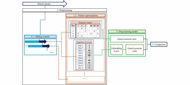
27. Exploring the Preprocessing of a Time-Series Administrative Healthcare Dataset on Deep Learning to Improve Prediction
Preprocessing methods are important in enhancing prediction performance for time-series administrative data. This study underscores the importance of preprocessing methods by comparing two data representation techniques, that can be used with sliding window techniques for time-series administrative healthcare data, for use with gated recurrent unit (GRU) networks. The first method uses a sequence event representation and the second employs a sequence matrix representation. The evaluation was conducted through a retrospective administrative healthcare data case study to predict multiple outcomes. The target outcomes were cases where a person’s First Healthcare Encounter indicated they were experiencing Homelessness (FHE-H) or involved Police (FHE-P), as recorded in the healthcare data. Results reveal that the GRU combined with sequence matrix representation and sliding window outperformed the sequence of events with the sliding window by over 20% in the area under the curve (AUC) and sensitivity for both outcomes. Given the data used in this analysis, sequence matrix representation was superior to sequence event representation while using GRU models. The results remained consistent across the evaluation in real-world clinical frameworks using two trigger methods for prediction time, such as the sliding window and clinical demand window structures.
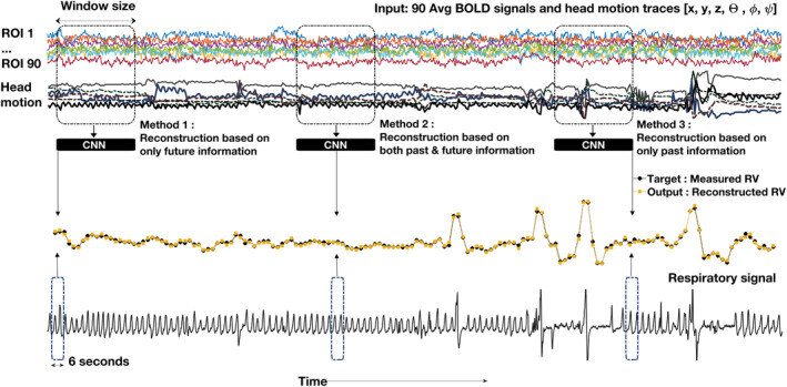
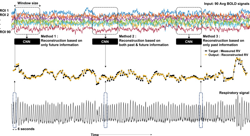
26. Machine Learning-based Estimation of Respiratory Fluctuations in a Healthy Adult Population using BOLD fMRI and Head Motion Parameters
Purpose External physiological monitoring is the primary approach to measure and remove effects of low‐frequency respiratory variation from BOLD‐fMRI signals. However, the acquisition of clean external respiratory data during fMRI is not always possible, so recent research has proposed using machine learning to directly estimate respiratory variation (RV), potentially obviating the need for external monitoring. In this study, we propose an extended method for reconstructing RV waveforms directly from resting state BOLD‐fMRI data in healthy adult participants with the inclusion of both BOLD signals and derived head motion parameters. Methods In the proposed method, 1D convolutional neural networks (1D‐CNNs) used BOLD signals and head motion parameters to reconstruct the RV waveform for the whole fMRI scan time. Resting‐state fMRI data and associated respiratory records from the Human Connectome Project in Young Adults (HCP‐YA) dataset are used to train and test the proposed method. Results Compared to using only BOLD‐fMRI data for a CNN input, this approach yielded improvements of 14% in mean absolute error, 24% in mean square error, 14% in correlation, and 12% in dynamic time warping. When tested on independent datasets, the method demonstrated generalizability, even in data with different TRs and physiological conditions. Conclusion This study shows that the respiratory variations could be reconstructed from BOLD‐fMRI data in the young adult population, and its accuracy could be improved using supportive data such as head motion parameters. The method also performed well on independent datasets with different experimental conditions.
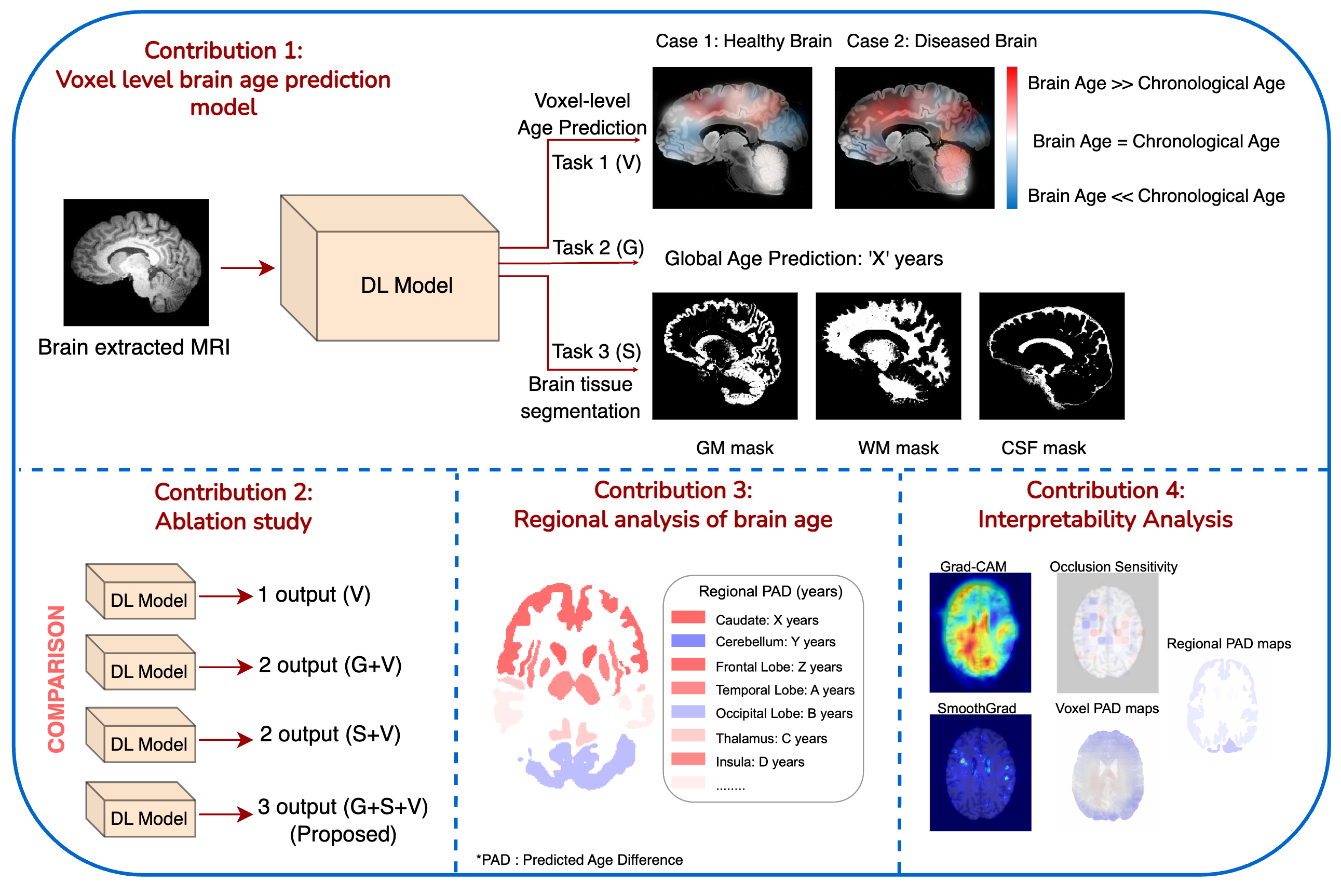
25. A Voxel-Level Approach to Brain Age Prediction: A Method to Assess Regional Brain Aging
Brain aging is a regional phenomenon, a facet that remains relatively under-explored within the realm of brain age prediction research using machine learning methods. Voxel-level predictions can provide localized brain age estimates that can provide granular insights into the regional aging processes. This is essential to understand the differences in aging trajectories in healthy versus diseased subjects. In this work, a deep learning-based multitask model is proposed for voxel-level brain age prediction from T1-weighted magnetic resonance images. The proposed model outperforms the models existing in the literature and yields valuable clinical insights when applied to both healthy and diseased populations. Regional analysis is performed on the voxel-level brain age predictions to understand aging trajectories of known anatomical regions in the brain and show that there exist disparities in regional aging trajectories of healthy subjects compared to ones with underlying neurological disorders such as Dementia and more specifically, Alzheimer’s disease.
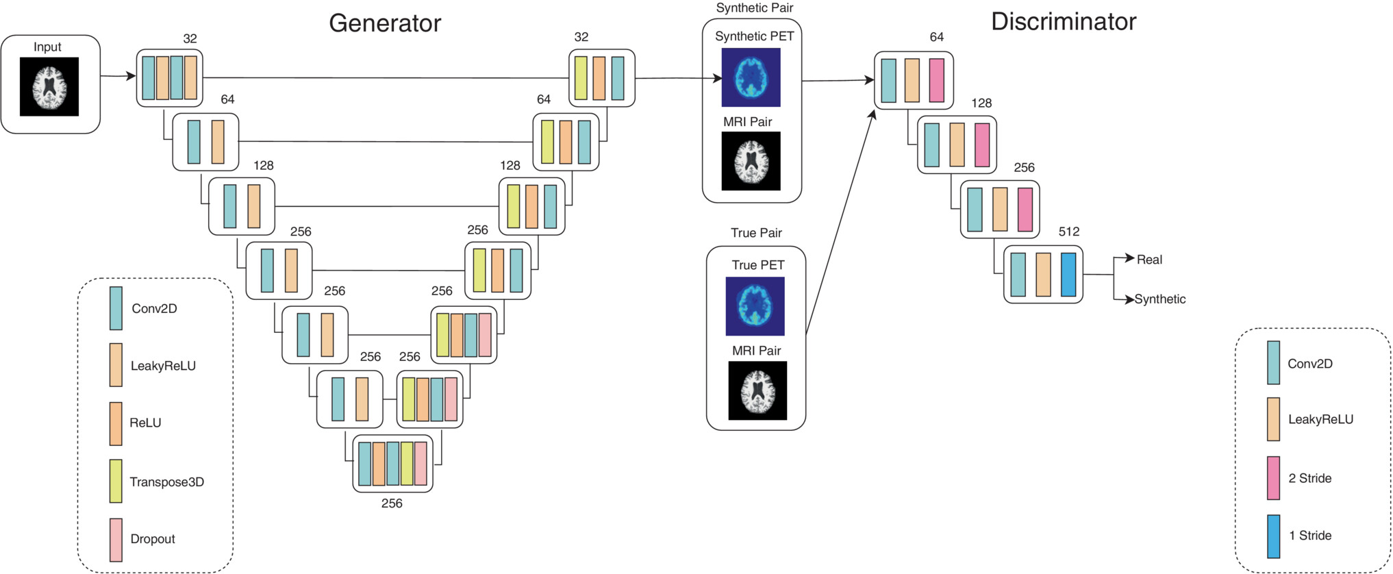
24. Image Translation for Estimating Two-Dimensional Axial Amyloid-Beta PET From Structural MRI
Amyloid-beta and brain atrophy are hallmarks for Alzheimer's Disease that can be targeted with positron emission tomography (PET) and MRI, respectively. MRI is cheaper, less-invasive, and more available than PET. There is a known relationship between amyloid-beta and brain atrophy, meaning PET images could be inferred from MRI. The purpose of this project is to build an image translation model using a Conditional Generative Adversarial Network able to synthesize Amyloid-beta PET images from structural MRI.
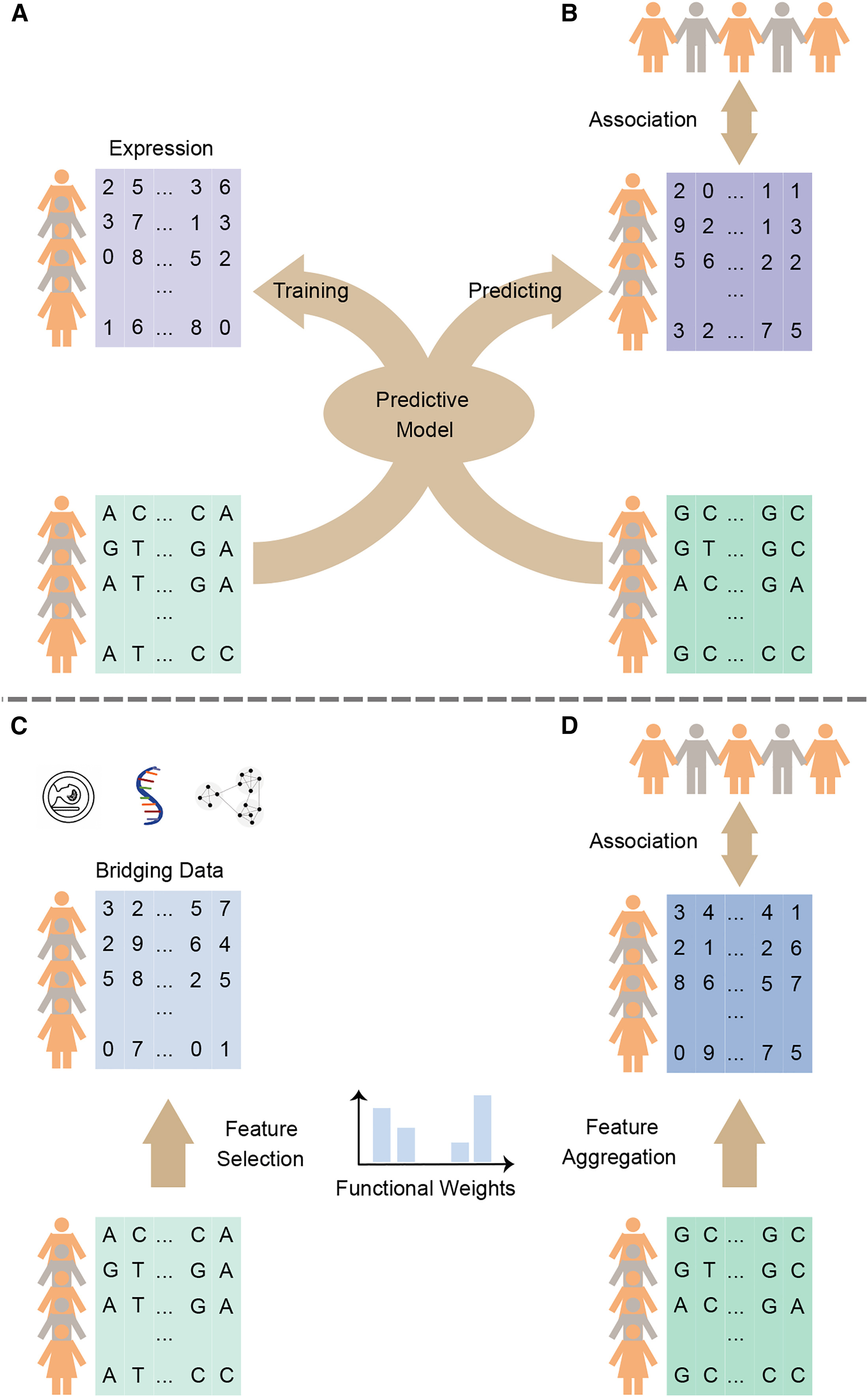
23. A Statistical Method for Image-Mediated Association Studies Discovers Genes and Pathways Associated with Four Brain Disorders
Brain imaging and genomics are critical tools enabling characterization of the genetic basis of brain disorders. However, imaging large cohorts is expensive and may be unavailable for legacy datasets used for genome-wide association studies (GWASs). Using an integrated feature selection/aggregation model, we developed an image-mediated association study (IMAS), which utilizes borrowed imaging/genomics data to conduct association mapping in legacy GWAS cohorts. By leveraging the UK Biobank image-derived phenotypes (IDPs), the IMAS discovered genetic bases underlying four neuropsychiatric disorders and verified them by analyzing annotations, pathways, and expression quantitative trait loci (eQTLs). A cerebellar-mediated mechanism was identified to be common to the four disorders. Simulations show that, if the goal is identifying genetic risk, our IMAS is more powerful than a hypothetical protocol in which the imaging results were available in the GWAS dataset. This implies the feasibility of reanalyzing legacy GWAS datasets without conducting additional imaging, yielding cost savings for integrated analysis of genetics and imaging.
22. Correspondence between BOLD fMRI task activation and cerebrovascular reactivity across the cerebral cortex
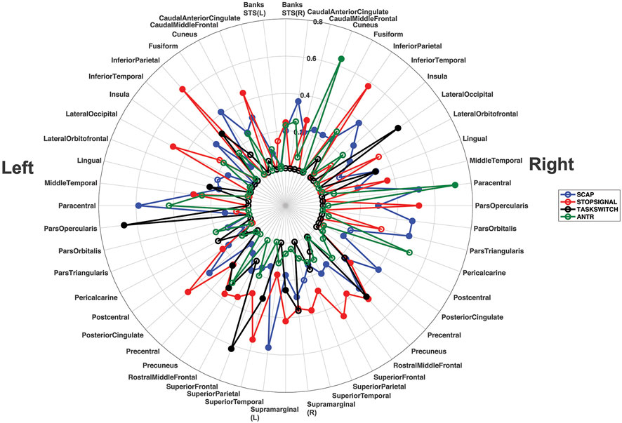
BOLD sensitivity to baseline perfusion and blood volume is a well-acknowledged fMRI confound. Vascular correction techniques based on cerebrovascular reactivity (CVR) might reduce variance due to baseline cerebral blood volume, however this is predicated on an invariant linear relationship between CVR and BOLD signal magnitude. Cognitive paradigms have relatively low signal, high variance and involve spatially heterogenous cortical regions; it is therefore unclear whether the BOLD response magnitude to complex paradigms can be predicted by CVR. The feasibility of predicting BOLD signal magnitude from CVR was explored in the present work across two experiments using different CVR approaches. The first utilized a large database containing breath-hold BOLD responses and 3 different cognitive tasks. The second experiment, in an independent sample, calculated CVR using the delivery of a fixed concentration of carbon dioxide and a different cognitive task. An atlas-based regression approach was implemented for both experiments to evaluate the shared variance between task-invoked BOLD responses and CVR across the cerebral cortex. Both experiments found significant relationships between CVR and task-based BOLD magnitude, with activation in the right cuneus and paracentral gyrus , and the left pars opercularis , superior frontal gyrus and inferior parietal cortex strongly predicted by CVR. The parietal regions bilaterally were highly consistent, with linear regressions significant in these regions for all four tasks. Group analyses showed that CVR correction increased BOLD sensitivity. Overall, this work suggests that BOLD signal response magnitudes to cognitive tasks are predicted by CVR across different regions of the cerebral cortex, providing support for the use of correction based on baseline vascular physiology.
21. Direct Machine Learning Reconstruction of Respiratory Variation Waveforms from Resting State fMRI Data in a Pediatric Population
In many functional magnetic resonance imaging (fMRI) studies, respiratory signals are unavailable or do not have acceptable quality due to issues with subject compliance, equipment failure or signal error. In large databases, such as the Human Connectome Projects, over half of the respiratory recordings may be unusable. As a result, the direct removal of low frequency respiratory variations from the blood oxygen level-dependent (BOLD) signal time series is not possible. This study proposes a deep learning-based method for reconstruction of respiratory variation (RV) waveforms directly from BOLD fMRI data in pediatric participants (aged 5 to 21 years old), and does not require any respiratory measurement device. To do this, the Lifespan Human Connectome Project in Development (HCP-D) dataset, which includes respiratory measurements, was used to both train a convolutional neural network (CNN) and evaluate its performance. Results show that a CNN can capture informative features from the BOLD signal time course and reconstruct accurate RV timeseries, especially when the subject has a prominent respiratory event. This work advances the use of direct estimation of physiological parameters from fMRI, which will eventually lead to reduced complexity and decrease the burden on participants because they may not be required to wear a respiratory bellows.
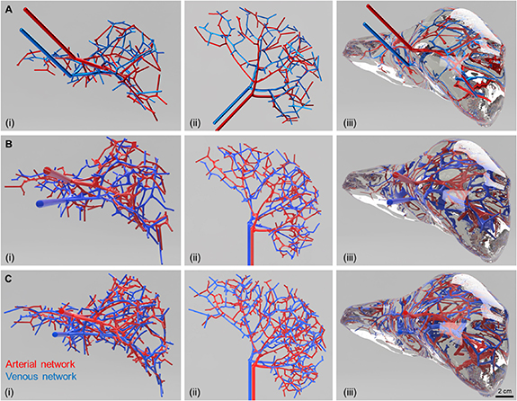
20. Closed-loop vasculature network design for bioprinting large, solid tissue scaffolds
Vascularization is an indispensable requirement for fabricating large solid tissues and organs. The natural vasculature derived from medical imaging modalities for large tissues and organs are highly complex and convoluted. However, the present bioprinting capabilities limit the fabrication of such complex natural vascular networks. Simplified bioprinted vascular networks, on the other hand, lack the capability to sustain large solid tissues. This work proposes a generalized and adaptable numerical model to design the vasculature by utilizing the tissue/organ anatomy. Starting with processing the patient's medical images, organ structure, tissue-specific cues, and key vasculature tethers are determined. An open-source abdomen magnetic resonance image dataset was used in this work. The extracted properties and cues are then used in a mathematical model for guiding the vascular network formation comprising arterial and venous networks. Next, the generated three-dimensional networks are used to simulate the nutrient transport and consumption within the organ over time and the regions deprived of the nutrients are identified. These regions provide cues to evolve and optimize the vasculature in an iterative manner to ensure the availability of the nutrient transport throughout the bioprinted scaffolds. The mass transport of six components of cell culture media—glucose, glycine, glutamine, riboflavin, human serum albumin, and oxygen was studied within the organ with designed vasculature. As the vascular structure underwent iterations, the organ regions deprived of these key components decreased significantly highlighting the increase in structural complexity and efficacy of the designed vasculature. The numerical method presented in this work offers a valuable tool for designing vascular scaffolds to guide the cell growth and maturation of the bioprinted tissues for faster regeneration post bioprinting.
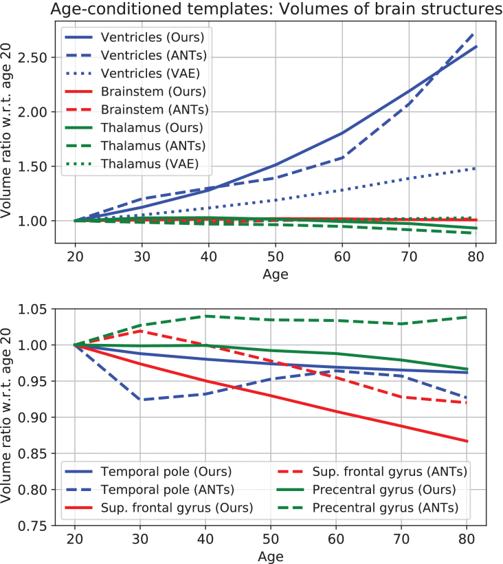
19. A Bidirectional Normalizing Flow Model of Brain Aging
Many machine learning tasks in neuroimaging aim at modeling complex relationships between a brain’s morphology as seen in structural MR images and clinical scores and variables of interest. A frequently modeled process is healthy brain aging for which many image-based brain age estimation or age-conditioned brain morphology template generation approaches exist. While age estimation is a regression task, template generation is related to generative modeling. Both tasks can be seen as inverse directions of the same relationship between brain morphology and age. However, this view is rarely exploited and most existing approaches train separate models for each direction. In this paper, we propose a novel bidirectional approach that unifies score regression and generative morphology modeling and we use it to build a bidirectional brain aging model. We achieve this by defining an invertible normalizing flow architecture that learns a probability distribution of 3D brain morphology conditioned on age. The use of full 3D brain data is achieved by deriving a manifold-constrained formulation that models morphology variations within a low-dimensional subspace of diffeomorphic transformations. This modeling idea is evaluated on a database of MR scans of more than 5000 subjects. The evaluation results show that our bidirectional brain aging model (1) accurately estimates brain age, (2) is able to visually explain its decisions through attribution maps and counterfactuals, (3) generates realistic age-specific brain morphology templates, (4) supports the analysis of morphological variations, and (5) can be utilized for subject-specific brain aging simulation.
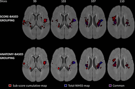
18. Lesion-symptom mapping with NIHSS sub-scores in ischemic stroke patients
Background: Lesion-symptom mapping (LSM) is a statistical technique to investigate the population-specific relationship between structural integrity and post-stroke clinical outcome. In clinical practice, patients are commonly evaluated using the National Institutes of Health Stroke Scale (NIHSS), an 11-domain clinical score to quantitate neurological deficits due to stroke. So far, LSM studies have mostly used the total NIHSS score for analysis, which might not uncover subtle structure–function relationships associated with the specific sub-domains of the NIHSS evaluation. Thus, the aim of this work was to investigate the feasibility to perform LSM analyses with sub-score information to reveal category-specific structure–function relationships that a total score may not reveal.Methods: Employing a multivariate technique, LSM analyses were conducted using a sample of 180 patients with NIHSS assessment at 48-hour post-stroke from the ESCAPE trial. The NIHSS domains were grouped into six categories using two schemes. LSM was conducted for each category of the two groupings and the total NIHSS score.Results:Sub-score LSMs not only identify most of the brain regions that are identified as critical by the total NIHSS score but also reveal additional brain regions critical to each function category of the NIHSS assessment without requiring extensive, specialised assessments.Conclusion:These findings show that widely available sub-scores of clinical outcome assessments can be used to investigate more specific structure–function relationships, which may improve predictive modelling of stroke outcomes in the context of modern clinical stroke assessments and neuroimaging.
17. Magnetic Resonance Imaging of Healthy Brain Aging: A Review
We present a review of the characterization of healthy brain aging using MRI with an emphasis on morphology, lesions, and quantitative MR parameters. A scope review found 6612 articles encompassing the keywords “Brain Aging” and “Magnetic Resonance”; papers involving functional MRI or not involving imaging of healthy human brain aging were discarded, leaving 2246 articles. We first consider some of the biogerontological mechanisms of aging, and the consequences of aging in terms of cognition and onset of disease. Morphological changes with aging are reviewed for the whole brain, cerebral cortex, white matter, subcortical gray matter, and other individual structures. In general, volume and cortical thickness decline with age, beginning in mid-life. Prevalent silent lesions such as white matter hyperintensities, microbleeds, and lacunar infarcts are also observed with increasing frequency. The literature regarding quantitative MR parameter changes includes T1, T2, T2*, magnetic susceptibility, spectroscopy, magnetization transfer, diffusion, and blood flow. We summarize the findings on how each of these parameters varies with aging. Finally, we examine how the aforementioned techniques have been used for age prediction. While relatively large in scope, we present a comprehensive review that should provide the reader with sound understanding of what MRI has been able to tell us about how the healthy brain ages.
16. The relationship between cognition and cerebrovascular reactivity: Implications for cognitive fMRI
Elucidating the brain regions and networks associated with cognitive processes has been the mainstay of task-based fMRI, under the assumption that BOLD signals are uncompromised by vascular function. This is despite the plethora of research highlighting BOLD modulations due to vascular changes induced by disease, drugs, and aging. On the other hand, BOLD fMRI-based assessment of cerebrovascular reactivity (CVR) is often used as an indicator of the brain's vascular health and has been shown to be strongly associated with cognitive function. This review paper considers the relationship between BOLD-based assessments of CVR, cognition and task-based fMRI. How the BOLD response reflects both CVR and neural activity, and how findings of altered CVR in disease and in normal physiology are associated with cognition and BOLD signal changes are discussed. These are pertinent considerations for fMRI applications aiming to understand the biological basis of cognition. Therefore, a discussion of how the acquisition of BOLD-based CVR can enhance our ability to map human brain function, with limitations and potential future directions, is presented.
15. Supervised machine learning tools: a tutorial for clinicians
In an increasingly data-driven world, artificial intelligence is expected to be a key tool for converting big data into tangible benefits and the healthcare domain is no exception to this. Machine learning aims to identify complex patterns in multi-dimensional data and use these uncovered patterns to classify new unseen cases or make data-driven predictions. In recent years, deep neural networks have shown to be capable of producing results that considerably exceed those of conventional machine learning methods for various classification and regression tasks. In this paper, we provide an accessible tutorial of the most important supervised machine learning concepts and methods, including deep learning, which are potentially the most relevant for the medical domain. We aim to take some of the mystery out of machine learning and depict how machine learning models can be useful for medical applications. Finally, this tutorial provides a few practical suggestions for how to properly design a machine learning model for a generic medical problem.
14. The Effect of Aging on Cerebral Blood Flow and Cortical Thickness with Application to Age Prediction
Cerebral cortex thinning and cerebral blood flow (CBF) reduction are typically observed during normal healthy aging. However, imaging-based age prediction models have primarily used morphological features of the brain. Complementary physiological CBF information might result in an improvement in age estimation. In this study, T1-weighted structural magnetic resonance imaging and arterial spin labeling CBF images were acquired in 146 healthy participants across the adult life span. Sixty-eight cerebral cortex regions were segmented, and the cortical thickness and mean CBF were computed for each region. Linear regression with age was computed for each region and data type, and laterality and correlation matrices were computed. Sixteen predictive models were trained with the cortical thickness and CBF data alone as well as a combination of both data types. The age explained more variance in the cortical thickness data than in the CBF data . All 16 models performed significantly better when combining both measurement types and using feature selection, and thus, we conclude that the inclusion of CBF data marginally improves age estimation.

13. High-resolution T2-FLAIR and non-contrast CT brain atlas of the elderly
Normative brain atlases are a standard tool for neuroscience research and are, for example, used for spatial normalization of image datasets prior to voxel-based analyses of brain morphology and function. Although many different atlases are publicly available, they are usually biased with respect to an imaging modality and the age distribution. Both effects are well known to negatively impact the accuracy and reliability of the spatial normalization process using non-linear image registration methods. An important and very active neuroscience area that lacks appropriate atlases is lesion- related research in elderly populations (e.g. stroke, multiple sclerosis) for which FLAIR MRI and non- contrast CT are often the clinical imaging modalities of choice. To overcome the lack of atlases for these tasks and modalities, this paper presents high-resolution, age-specific FLAIR and non-contrast CT atlases of the elderly generated using clinical images.
12. Interdatabase Variability of Cortical Thickness Measurements
The phenomenon of cortical thinning with age has been well established; however, the measured rate of change varies between studies. The source of this variation could be image acquisition techniques including hardware and vendor specific differences. Databases are often consolidated to increase the number of subjects but underlying differences between these datasets could have undesired effects. We explore differences in cerebral cortex thinning between 4 databases, totaling 1382 subjects. We investigate several aspects of these databases, including: 1) differences between databases of cortical thinning rates versus age, 2) correlation of cortical thinning rates between regions for each database, and 3) regression bootstrapping to determine the effect of the number of subjects included. We also examined the effect of different databases on age prediction modeling. Cortical thinning rates were significantly different between databases in all 68 parcellated regions (ANCOVA, P < 0.001). Subtle differences were observed in correlation matrices and bootstrapping convergence. Age prediction modeling using a leave-one-out cross-validation approach showed varying prediction performance between databases. When a database was used to calibrate the model and then applied to another database, prediction performance consistently decreased. We conclude that there are indeed differences in the measured cortical thinning rates between these large-scale databases.
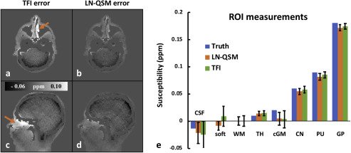
11. Whole Head Quantitative Susceptibility Mapping Using a Least-norm Direct Dipole Inversion Method
A new dipole field inversion method for whole head quantitative susceptibility mapping (QSM) is proposed. Instead of performing background field removal and local field inversion sequentially, the proposed method performs dipole field inversion directly on the total field map in a single step. To aid this under-determined and ill-posed inversion process and obtain robust QSM images, Tikhonov regularization is implemented to seek the local susceptibility solution with the least-norm (LN) using the L-curve criterion. The proposed LN-QSM does not require brain edge erosion, thereby preserving the cerebral cortex in the final images. This should improve its applicability for QSM-based cortical grey matter measurement, functional imaging and venography of full brain. Furthermore, LN-QSM also enables susceptibility mapping of the entire head without the need for brain extraction, which makes QSM reconstruction more automated and less dependent on intermediate pre-processing methods and their associated parameters. It is shown that the proposed LN-QSM method reduced errors in a numerical phantom simulation, improved accuracy in a gadolinium phantom experiment, and suppressed artefacts in nine subjects, as compared to two-step and other single-step QSM methods. Measurements of deep grey matter and skull susceptibilities from LN-QSM are consistent with established reconstruction methods.
10. Modelling Hyperoxia-induced BOLD Signal Dynamics to Estimate Cerebral Blood Flow, Volume and Mean Transit Time
A new method is proposed for obtaining cerebral perfusion measurements whereby blood oxygen level dependent (BOLD) MRI is used to dynamically monitor hyperoxia-induced changes in the concentration of deoxygenated hemoglobin in the cerebral vasculature. The data is processed using kinetic modeling to yield perfusion metrics, namely: cerebral blood flow (CBF), cerebral blood volume (CBV), and mean transit time (MTT). Ten healthy human subjects were continuously imaged with BOLD sequence while a hyperoxic (70% O2) state was interspersed with baseline periods of normoxia. The BOLD time courses were fit with exponential uptake and decay curves and a biophysical model of the BOLD signal was used to estimate oxygen concentration functions. The arterial input function was derived from end-tidal oxygen measurements, and a deconvolution operation between the tissue and arterial concentration functions was used to yield CBF. The venous component of the CBV was calculated from the ratio of the integrals of the estimated tissue and arterial concentration functions. We conclude that it is possible to derive CBF, CBV and MTT metrics within expected physiological ranges via analysis of dynamic BOLD fMRI acquired during a period of hyperoxia.

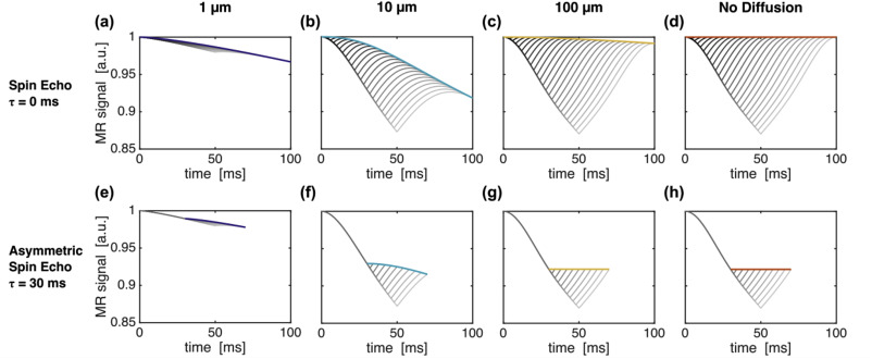
9. Gas-free Calibrated fMRI with a Correction for Vessel-Size Sensitivity
Calibrated functional magnetic resonance imaging (fMRI) is a method to independently measure the metabolic and hemodynamic contributions to the blood oxygenation level dependent (BOLD) signal. This technique typically requires the use of a respiratory challenge, such as hypercapnia or hyperoxia, to estimate the calibration constant, M. There has been a recent push to eliminate the gas challenge from the calibration procedure using asymmetric spin echo (ASE) based techniques. This study uses simulations to better understand spin echo (SE) and ASE signals, analytical modelling to characterize the signal evolution, and in vivo imaging to validate the modelling. Using simulations, it is shown how ASE imaging generally underestimates M and how this depends on several parameters of the acquisition, including echo time and ASE offset, as well as the vessel size. This underestimation is the result of imperfect SE refocusing due to diffusion of water through the extravascular environment surrounding the microvasculature. By empirically characterizing this SE attenuation as an exponential decay that increases with echo time, we have proposed a quadratic ASE biophysical signal model. This model allows for the characterization and compensation of the SE attenuation if SE and ASE signals are acquired at multiple echo times. This was tested in healthy subjects and was found to significantly increase the estimates of M across grey matter. These findings show promise for improved gas-free calibration and can be extended to other relaxation-based imaging studies of brain physiology.
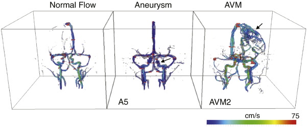
8. Dynamic Phase Contrast Magnetic Resonance Imaging for the Assessment of Arteriovenious Malformation and Aneurysm Pressure
Purpose: To explore phase contrast (PC) magnetic resonance imaging of aneurysms and arteriovenous malformations (AVM). PC imaging obtains a vector field of the velocity and can yield additional hemodynamic information, including: volume flow rate (VFR) and intravascular pressure. We expect to find lower VFR distal to aneurysms and higher VFR in vessels of the AVM. Materials and Methods: Five cerebral aneurysm and three AVM patients were imaged with PC techniques and compared to VFR of a healthy cohort. VFR was calculated in vessel segments in each patient and compared statistically to the healthy cohort by computing the z-score. Intravascular pressure was calculated in the aneurysms and in the nidus of each AVM. Results: We found that patients with aneurysm had z < −0.48 in vessels distal to the aneurysm (reduced flow), while AVM patients had z > 6 in some vessels supplying and draining the nidus (increased flow). Pressures in aneurysms were highly variable between subjects and location, while in the nidus of the AVM patients; pressure trended higher in larger AVMs. Conclusion: The study findings confirm the expectation of lower distal flow in aneurysm and higher arterial and venous flow in AVM patients.
7. Phase Error Correction in Time-Averaged 3D Phase Contrast Magnetic Resonance Imaging of the Cerebral Vasculature
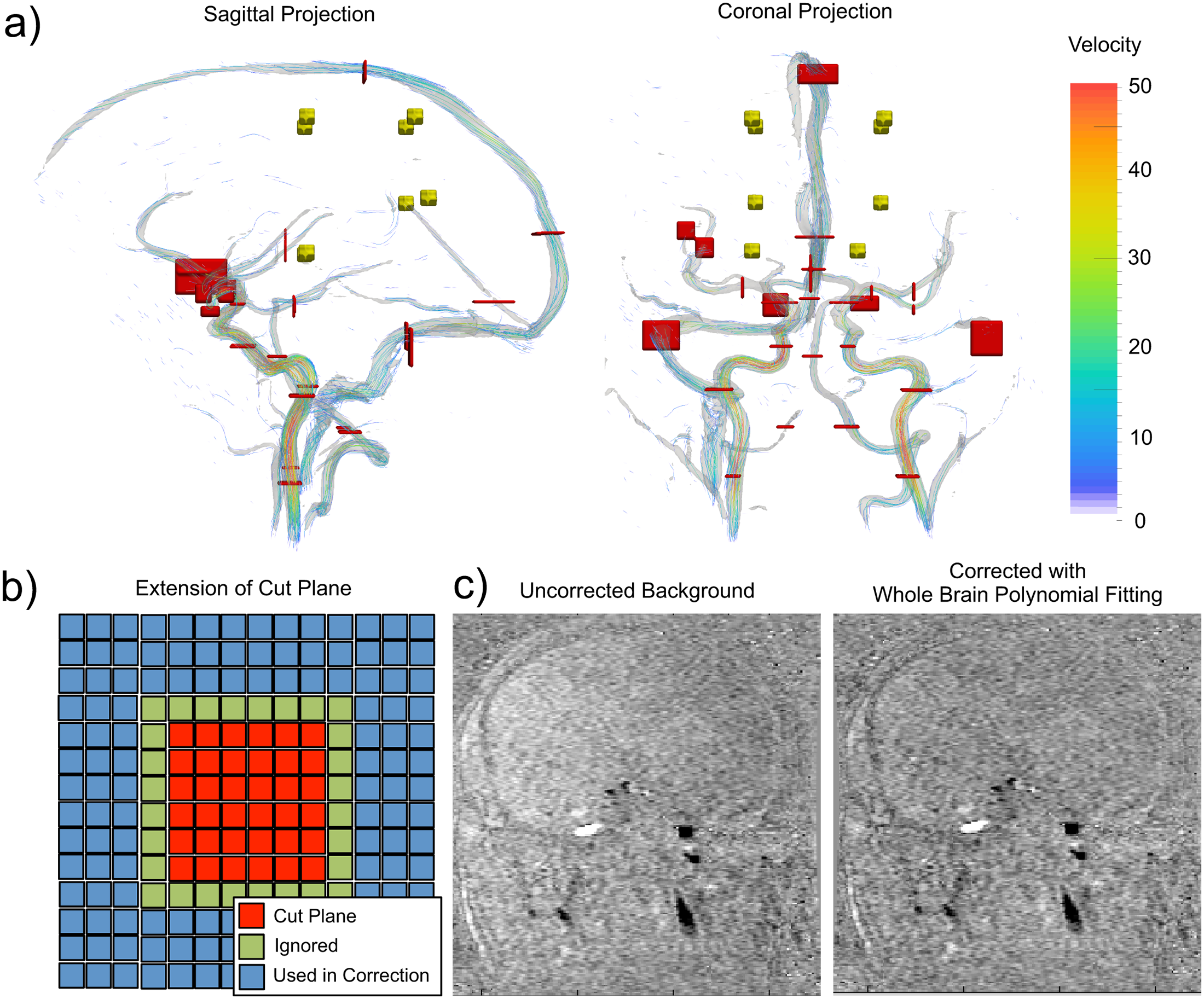
Purpose:Volume flow rate (VFR) measurements based on phase contrast (PC)-magnetic resonance (MR) imaging datasets have spatially varying bias due to eddy current induced phase errors. The purpose of this study was to assess the impact of phase errors in time averaged PC-MR imaging of the cerebral vasculature and explore the effects of three common correction schemes (local bias correction (LBC), local polynomial correction (LPC), and whole brain polynomial correction (WBPC)).Methods:Measurements of the eddy current induced phase error from a static phantom were first obtained. In thirty healthy human subjects, the methods were then assessed in background tissue to determine if local phase offsets could be removed. Finally, the techniques were used to correct VFR measurements in cerebral vessels and compared statistically.Results:In the phantom, phase error was measured to be <2.1 ml/s per pixel and the bias was reduced with the correction schemes. In background tissue, the bias was significantly reduced, by 65.6% (LBC), 58.4% (LPC) and 47.7% (WBPC) (p < 0.001 across all schemes). Correction did not lead to significantly different VFR measurements in the vessels (p = 0.997). In the vessel measurements, the three correction schemes led to flow measurement differences of -0.04 ± 0.05 ml/s, 0.09 ± 0.16 ml/s, and -0.02 ± 0.06 ml/s. Although there was an improvement in background measurements with correction, there was no statistical difference between the three correction schemes (p = 0.242 in background and p = 0.738 in vessels).Conclusions:While eddy current induced phase errors can vary between hardware and sequence configurations, our results showed that the impact is small in a typical brain PC-MR protocol and does not have a significant effect on VFR measurements in cerebral vessels.
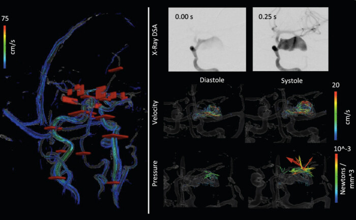
6. Flow and Hemodynamic Alteration in a Giant Cerebral Aneurysm Treated with a Pipeline Stent
Many risk factors have been proposed in the development of cerebral aneurysms. Hemodynamics including blood velocity, volume flow rate (VFR), and intravascular pressure are thought to be prognostic indicators of aneurysm development. We hypothesize that treatment of a cerebral aneurysm using a flow-diverting stent will bring these hemodynamic parameters closer to those observed on the contralateral side. In the current study, a patient with a giant cerebral aneurysm was studied pre- and postoperatively using phase contrast MRI (PC-MRI) to measure the hemodynamic changes resulting from the deployment of a flow-diverting stent. PC-MRI was used to calculate intravascular pressure, which was compared to more invasive endovascular catheter-derived measurements. After stent placement, the measured VFRs in vessels of the treated hemisphere approached those measured on the contralateral side, and flow symmetry changed from a laterality index of -0.153 to 0.116 in the middle cerebral artery. Pressure estimates derived from the PC-MRI velocity data had an average difference of 6.1% compared to invasive catheter transducer measurements. PC-MRI can measure the hemodynamic parameters with the same accuracy as invasive methods pre- and postoperatively.
5. Cerebrovascular Magnetic Resonance Imaging: A Review of State-of-the-Art Approaches, Methods and Techniques
Cerebrovascular imaging is of great interest in the understanding of neurological disease. MRI is a non‐invasive technology that can visualize and provide information on: (i) the structure of major blood vessels; (ii) the blood flow velocity in these vessels; and (iii) the microcirculation, including the assessment of brain perfusion. Although other medical imaging modalities can also interrogate the cerebrovascular system, MR provides a comprehensive assessment, as it can acquire many different structural and functional image contrasts whilst maintaining a high level of patient comfort and acceptance. The extent of examination is limited only by the practicalities of patient tolerance or clinical scheduling limitations. Currently, MRI methods can provide a range of metrics related to the cerebral vasculature, including: (i) major vessel anatomy via time‐of‐flight and contrast‐enhanced imaging; (ii) blood flow velocity via phase contrast imaging; (iii) major vessel anatomy and tissue perfusion via arterial spin labeling and dynamic bolus passage approaches; and (iv) venography via susceptibility‐based imaging. When designing an MRI protocol for patients with suspected cerebral vascular abnormalities, it is appropriate to have a complete understanding of when to use each of the available techniques in the ‘MR angiography toolkit’. In this review article, we: (i) overview the relevant anatomy, common pathologies and alternative imaging modalities; (ii) describe the physical principles and implementations of the above listed methods; (iii) provide guidance on the selection of acquisition parameters; and (iv) describe the existing and potential applications of MRI to the cerebral vasculature and diseases. The focus of this review is on obtaining an understanding through the application of advanced MRI methodology of both normal and abnormal blood flow in the cerebrovascular arteries, capillaries and veins.
4. Phase Contrast MR Imaging Measurements of Blood Flow in Healthy Human Cerebral Vessel Segments
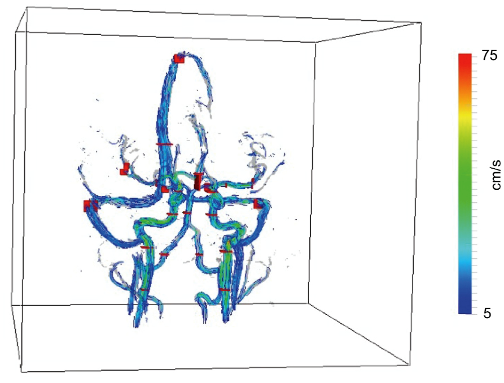
Phase contrast (PC) magnetic resonance imaging was used to obtain velocity measurements in 30 healthy subjects to provide an assessment of hemodynamic parameters in cerebral vessels. We expect a lower coefficient-of-variation (COV) of the volume flow rate (VFR) compared to peak velocity (Vpeak) measurements and the COV to increase in smaller caliber arteries compared to large arteries.PC velocity maps were processed to calculate Vpeak and VFR in 26 vessel segments. The mean, standard deviation and COV, of Vpeak and VFR in each segment were calculated. A bootstrap-style analysis was used to determine the minimum number of subjects required to accurately represent the population. Significance of Vpeak and VFR asymmetry was assessed in 10 vessel pairs.The bootstrap analysis suggested that averaging more than 20 subjects would give consistent results. When averaged over the subjects, Vpeak and VFR ranged from 5.2 ± 7.1 cm/s, 0.41 ± 0.58 ml/s (in the anterior communicating artery; mean ± standard deviation) to 73 ± 23 cm/s, 7.6 ± 1.7 ml/s (in the left internal carotid artery), respectively. A tendency for VFR to be higher in the left hemisphere was observed in 88.8% of artery pairs, while the VFR in the right transverse sinus was larger. The VFR COV was larger than Vpeak COV in 57.7% of segments, while smaller vessels had higher COV.Significance and potential impact: VFR COV was not generally higher than Vpeak COV. COV was higher in smaller vessels as expected. These summarized values provide a base against which Vpeak and VFR in various disease states can be compared.
3. Accelerated Passive MR Catheter Tracking into the Carotid Artery of Canines Magnetic Resonance Imaging
Background:Using magnetic resonance (MR) imaging for navigating catheters has several advantages when compared with the current “gold standard” modality of X-ray imaging. A significant drawback to interventional MR is inferior temporal and spatial resolutions, as high spatial resolution images cannot be collected and displayed at rates equal to X-ray imaging. In particular, passive MR catheter tracking experiments that use positive contrast mechanisms have poor temporal imaging rates and signal-to-noise ratio. As a result, with passive methods, it is often difficult to reconstruct motion artifact-free tracking images from areas with motion, such as the thoracic cavity.Methods:In this study, several accelerated MR acquisition strategies, including parallel imaging and compressed sensing (CS), were evaluated to determine which method is most effective at improving the frame rate and passive detection of catheters in regions of physiological motion. Device navigation was performed both in vitro, through the aortic arch of an anthropomorphic chest phantom, and in vivo from the femoral artery, up the descending aorta into the supra-aortic branching vessels in canines.Results and Discussion:The different parallel imaging methods produced images of low quality. CS with a two-fold acceleration was found to be the most effective method for generating tracking images, improving the image frame rate to 5.2 Hz, while maintaining a relatively high in-plane resolution. Using CS, motion artifact was decreased and the catheters were visualized with good conspicuity near the heart.Conclusions:The improvement in the imaging frame rate by image acceleration was sufficient to overcome motion artifacts and to better visualize catheters in the thoracic cavity with passive tracking. CS preformed best at tracking. Navigation with passive MR catheter tracking was demonstrated from the femoral artery to the carotid artery in canines.
2. Closed-Loop Circulation Phantom with Heart and Lung Motion for Validating Passive Magnetic Resonance Catheter Tracking
Purpose:To develop an anthropomorphic phantom to simulate heart, lung, and blood motion. Magnetic resonance imaging (MRI) is sensitive to image distortion and artifacts caused by motion. Imaging phantoms are used to test new sequences, but generally, these phantoms lack physiological motion. For the validation of new MR-based endovascular interventional and other techniques, we developed a dynamic motion phantom that is suitable for initial in vitro and more realistic validation studies that should occur before animal experiments.Materials and Methods:An anthropomorphic phantom was constructed to model the thoracic cavity, including respiratory and cardiac motions, and moving blood. Several MRI methods were used to validate the phantom performance: anatomical scanning, rapid temporal imaging, digital subtraction angiography, and endovascular tracking. The quality and nature of the motion artifact in these images were compared with in vivo images.Results:The closed-loop motion phantom correctly represented key features in the thorax, was MR-compatible, and was able to reproduce similar motion artifacts and effects as seen in in vivo images. The phantom provided enough physiological realism that it was able to ensure a suitable challenge in an in vitro catheter tracking experiment.Conclusion:A phantom was created and used for testing interventional catheter guiding. The images produced had similar qualities to those found in vivo. This phantom had a high degree of appropriate anthropomorphic and physiological qualities. Ethically, use of this phantom is highly appropriate when first testing new MRI techniques prior to conducting animal studies.

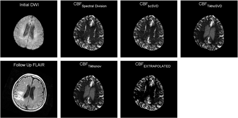
1. Deconvolution with Simple Extrapolation for Improved CBF Measurements in DSC-MRI during Acute Ischemic Stroke
Magnetic resonance (MR) perfusion imaging is a clinical technique for measuring brain blood flow parameters during stroke and other ischemic events. Ischemia in brain tissue can be difficult to accurately measure or visualize when using MR-derived cerebral blood flow (CBF) maps. The deconvolution techniques used to estimate flow can introduce a mean transit time-dependent bias following application of noise stabilization techniques. The underestimation of the CBF values, greatest in normal tissues, causes a decrease in the image contrast observed in CBF maps between normally perfused and ischemic tissues; resulting in ischemic areas becoming less conspicuous. Through application of the proposed simple extrapolation technique, CBF biases are reduced when missing high-frequency signal components in the MR data removed during deconvolution noise stabilization are restored. The extrapolation approach was compared with other methods and showed a statistically significant increase in image contrast in CBF maps between normal and ischemic tissues for white matter (P<.05) and performed better than most other methods for gray matter. Receiver operator characteristic curve analysis demonstrated that extrapolated CBF maps better-detected penumbral regions. Extrapolated CBF maps provided more accurate CBF estimates in simulations, suggesting that the approach may provide a better prediction of outcome in the absence of treatment.